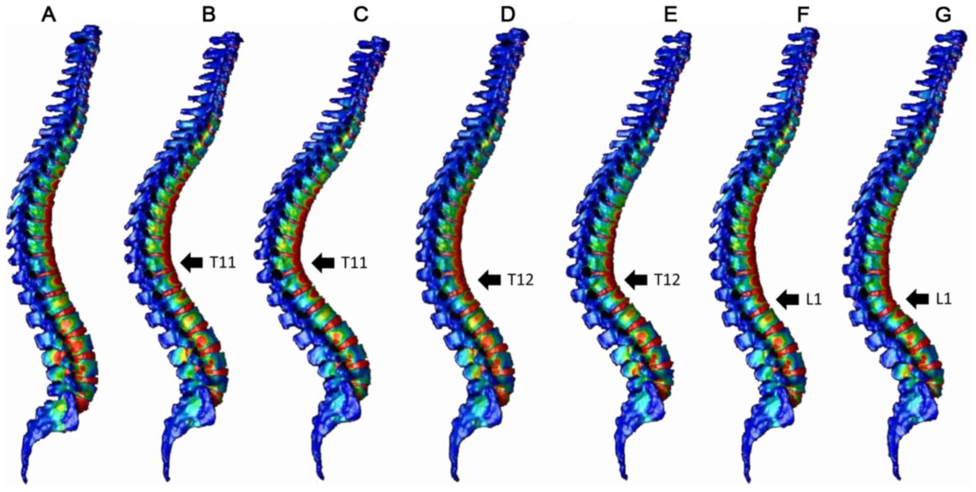
Vertebral fractures commonly occur at the thoracolumbar junction. These fractures can be treated with mild residual deformity in many cases, but are reportedly associated with increased risk of secondary vertebral fractures. In the present study, a three‑dimensional (3D) whole spine model was constructed using the finite element method to explore the mechanism of development of compression fractures. The 3D model of the whole spine, from the cervical spine to the pelvis, was constructed from computed tomography (CT) images of an adult male. Using a normal spine model and spine models with compression fractures at the T11, T12 or L1 vertebrae, the distribution of strain was analyzed in the vertebrae after load application. The normal spine model demonstrated greater strain around the thoracolumbar junction and the middle thoracic spine, while the compression fracture models indicated focused strain at the fracture site and adjacent vertebrae. Increased load time resulted in the extension of the strain region up to the middle thoracic spine. The present findings, that secondary vertebral fractures commonly occur around the fracture site, and may also affect the thoracic vertebrae, are consistent with previous clinical and experimental results. These results suggest that follow‑up examinations of compression fractures at the thoracolumbar junction should include the thoracic spine and adjacent vertebrae. The current data also demonstrate that models created from CT images can be used for various analyses.

PDF] Finite element analysis of compression fractures at the thoracolumbar junction using models constructed from medical images

Computer-Assisted Quantification
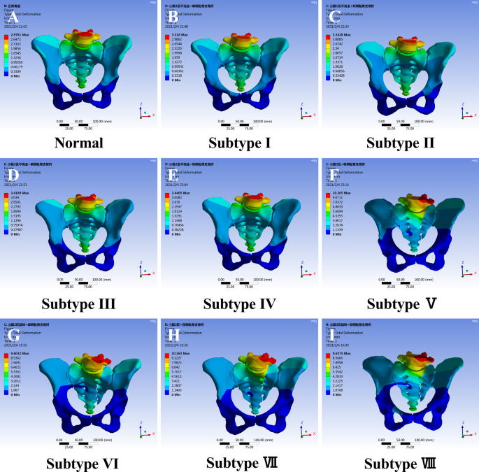
The morphological mapping of lateral compression type 1 pelvic fracture and pelvic ring stability classification: a finite element analysis, Journal of Orthopaedic Surgery and Research

3D patient-specific finite element models of the proximal femur based on DXA towards the classification of fracture and non-fracture cases - ScienceDirect

Applied Sciences, Free Full-Text
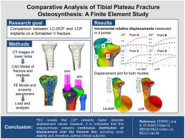
Comparative analysis of tibial plateau fracture osteosynthesis: A finite element study,Journal of the Mechanical Behavior of Biomedical Materials - X-MOL

PDF] Finite Element Method Analysis of Compression Fractures on Whole-Spine Models Including the Rib Cage

バイオメカニクス 山口大学大学院医学系研究科整形外科学
Lumbar Compression Fracture - Physiopedia
Frontiers Biomechanical Evaluation and the Assisted 3D Printed Model in the Patient-Specific Preoperative Planning for Thoracic Spinal Tuberculosis: A Finite Element Analysis

Biomechanics and finite element analysis of a novel plate designed for posterolateral tibial plateau fractures via the anterolateral approach

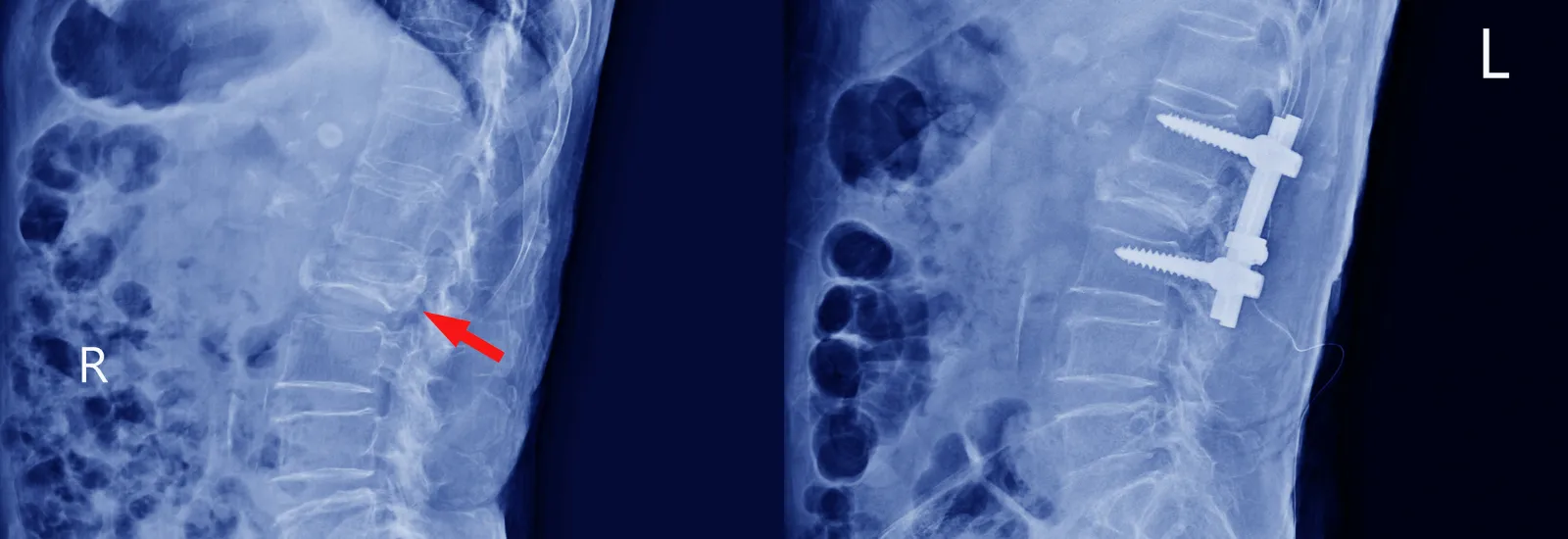
Vertebral body compression fracture - T12, Radiology Case
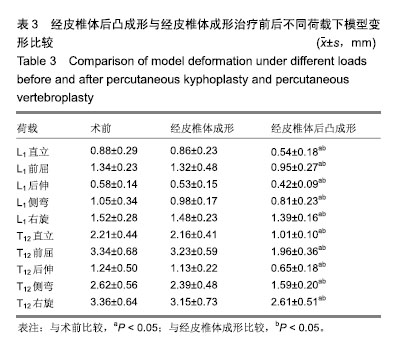
Mechanical changes of percutaneous kyphoplasty and percutaneous vertebroplasty in the treatment of thoracolumbar compressive fractures in three-dimensional vertebral models