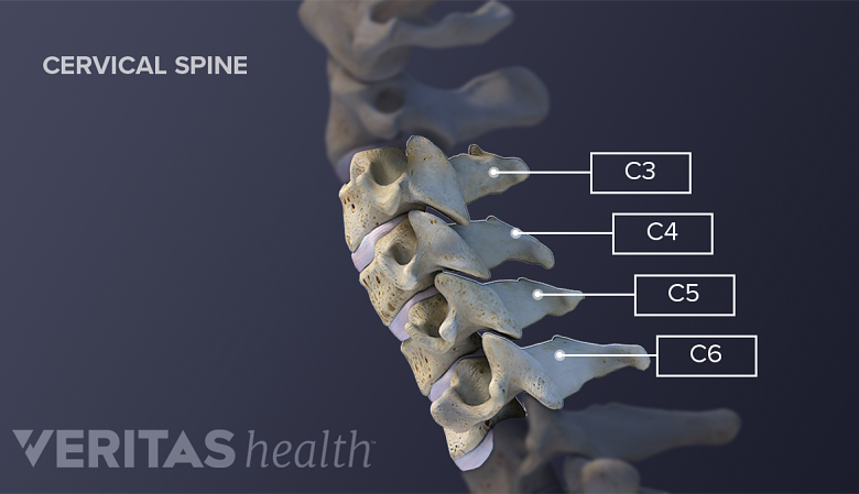
Of the seven cervical vertebrae, C3 through C6 have typical anatomy, while C7 looks very similar. C1 (atlas) and C2 (axis) have very distinct anatomical features. For a basic anatomic description o

Typical cervical vertebrae, Radiology Reference Article

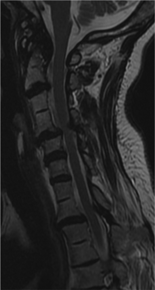
MRI of the cervical spine showing normal features.

Imaging features of the postoperative spine: a guide to basic understanding of spine surgical procedures, Insights into Imaging

Vertebral scalloping, Radiology Reference Article, Radiopaedia.org

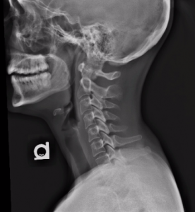
C-spine x-ray interpretation - Don't Forget the Bubbles

Cervical Spine Radiographic Anatomy –

Cervical radiographs taken one year before chiropractic attention. (A)
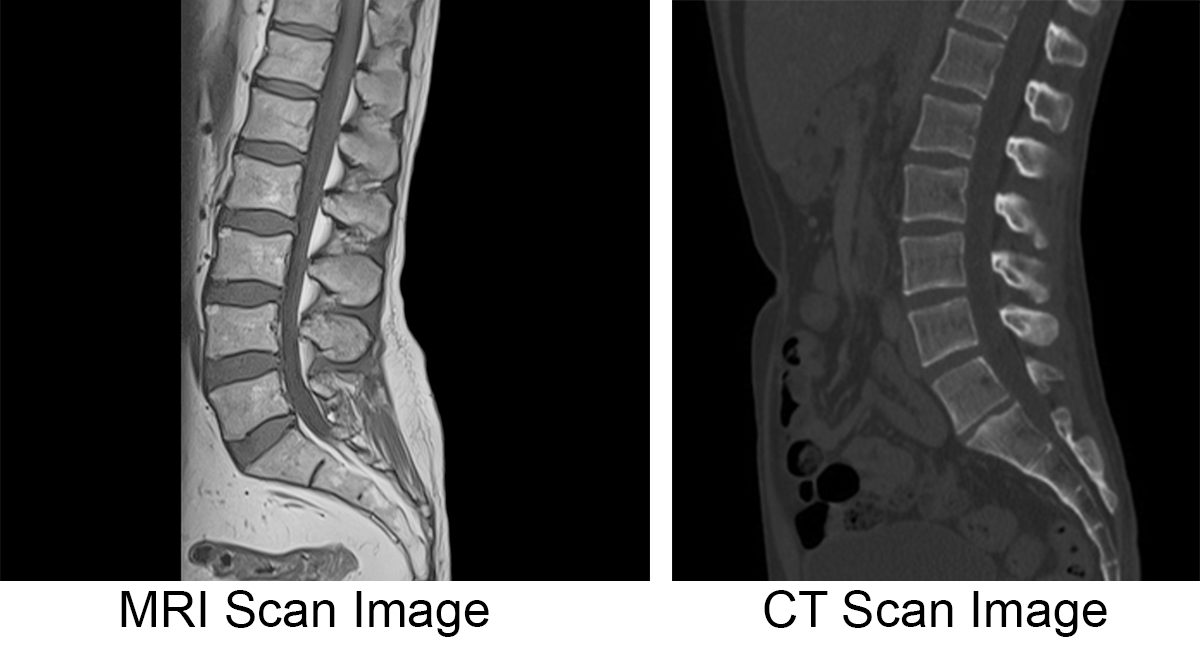
MRI vs. CT Scan; Diagnosing Spine & Neck Injuries & Degenerative Diseases

Small Animal Spinal Radiography Series: Cervical Spine Radiography
Part 9: Spine Radiology Key

Correlation and reliability of cervical sagittal alignment parameters between plain radiographs and multipositional MRI images

Typical cervical vertebrae, Radiology Reference Article
Head and Neck – Undergraduate Diagnostic Imaging Fundamentals

Cervical Spine – Anatomy, Diseases and Treatments
:max_bytes(150000):strip_icc()/GettyImages-1129624171-08e8d926acc944b697fc54acb2647660.jpg)