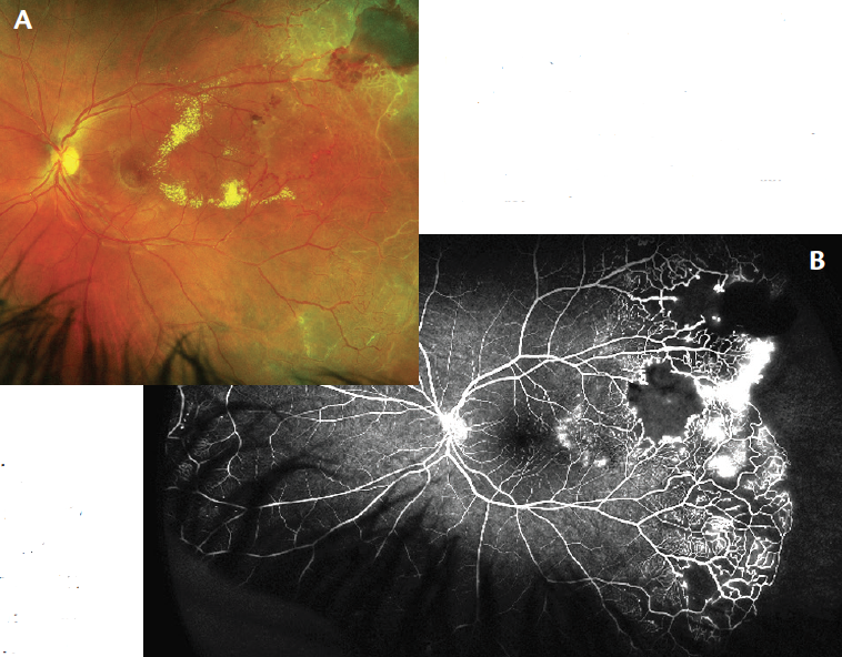
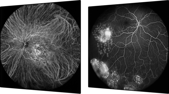
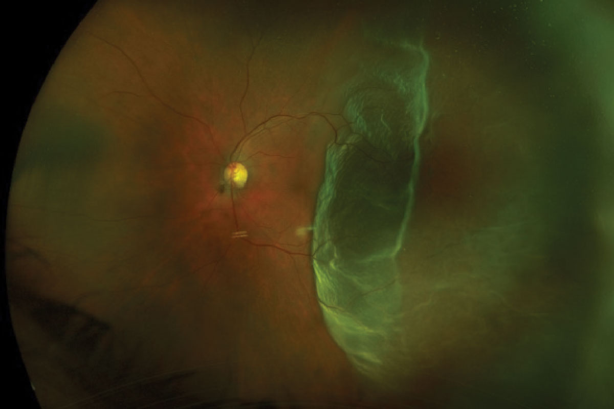
Download scientific diagram | Ultra-wide-field fundus photographs and ultra-wide-field fluorescein angiographic imaging of ocular toxocariasis. (A) A granuloma with mild vitreous opacity. (B) A tractional retinal fold with localized tractional retinal detachment. (C) Diffuse peripheral vascular leakage. (D) A prominent optic disc leakage. from publication: The Clinical Characteristics of Ocular Toxocariasis in Jeju Island Using Ultra-wide-field Fundus Photography | Toxocariasis, Ocular and Photography | ResearchGate, the professional network for scientists.
Ultra-Widefield Imaging Guides Coats Disease Treatment - Retina Today
SPECTRALIS Ultra-Widefield Angiography Module

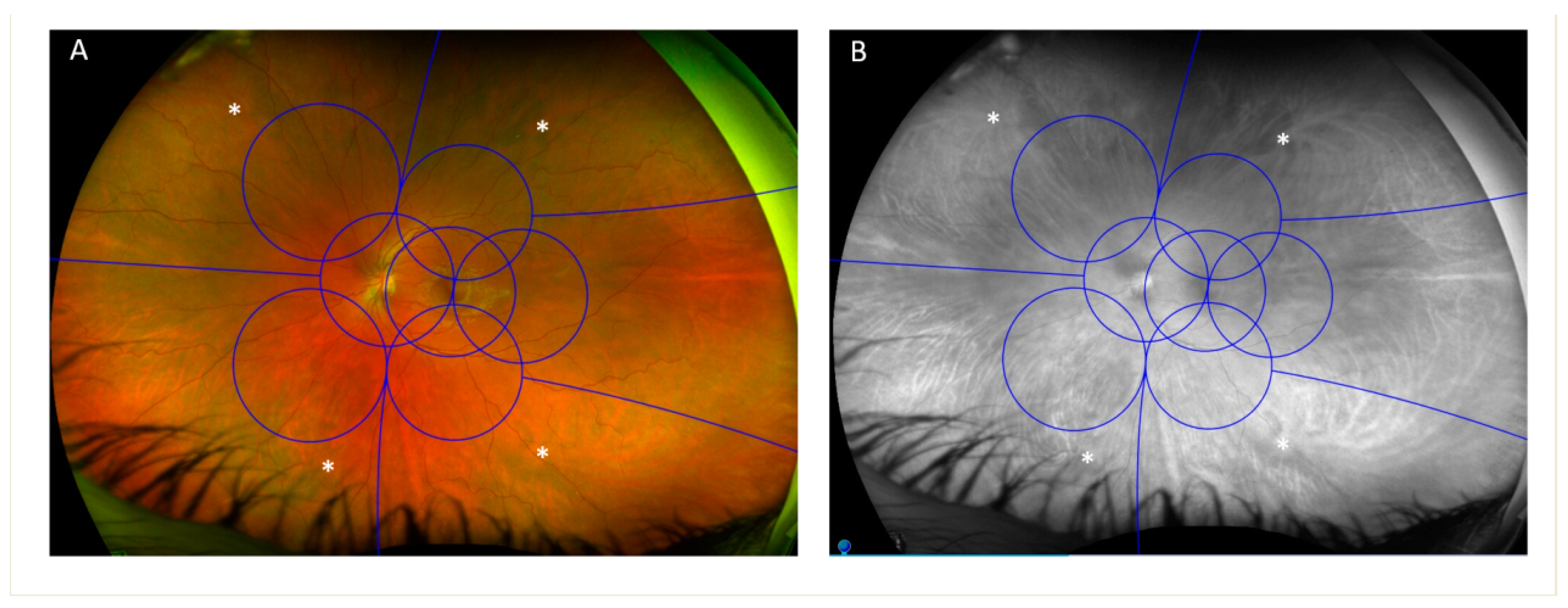
Ultra-wide-field fundus images with overlay of the Early Treatment

Jong Young Lee's research works Jeju National University Hospital, Jeju City and other places
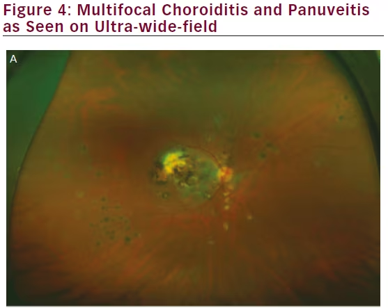
Wide-field Imaging of Retinal Diseases - touchOPHTHALMOLOGY

Comparison of true-colour wide-field confocal scanner imaging with standard fundus photography for diabetic retinopathy screening

Ultra-Widefield Imaging: Advancing the Understanding and Management of Diabetic Retinopathy - Retina Today

PDF) The Clinical Characteristics of Ocular Toxocariasis in Jeju Island Using Ultra-wide-field Fundus Photography

Ultra-wide-field imaging in diabetic retinopathy; an overview - ScienceDirect

Widefield and Ultra-widefield Imaging: When and Why to Use Them
Ultra-Widefield Imaging: Expand Your Horizons

Ultra-wide-field fundus photography compared to ophthalmoscopy in diagnosing and classifying major retinal diseases
JCM, Free Full-Text