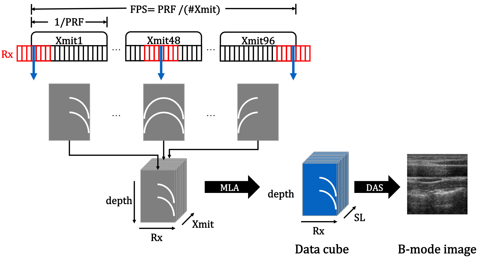
Download scientific diagram | Imaging flow of the standard B-mode ultrasound imaging. PRF : pulse repetition frequency, FPS : frame per second. from publication: Deep Learning in RF Sub-sampled B-mode Ultrasound Imaging | In portable, three dimensional, and ultra-fast ultrasound (US) imaging systems, there is an increasing need to reconstruct high quality images from a limited number of RF data from receiver (Rx) or scan-line (SC) sub-sampling. However, due to the severe side lobe artifacts | Radio Frequency, Ultrasound Imaging and Ultrasonography | ResearchGate, the professional network for scientists.

Ultrasound imaging of bone fractures, Insights into Imaging
1712.06096] Efficient B-mode Ultrasound Image Reconstruction from
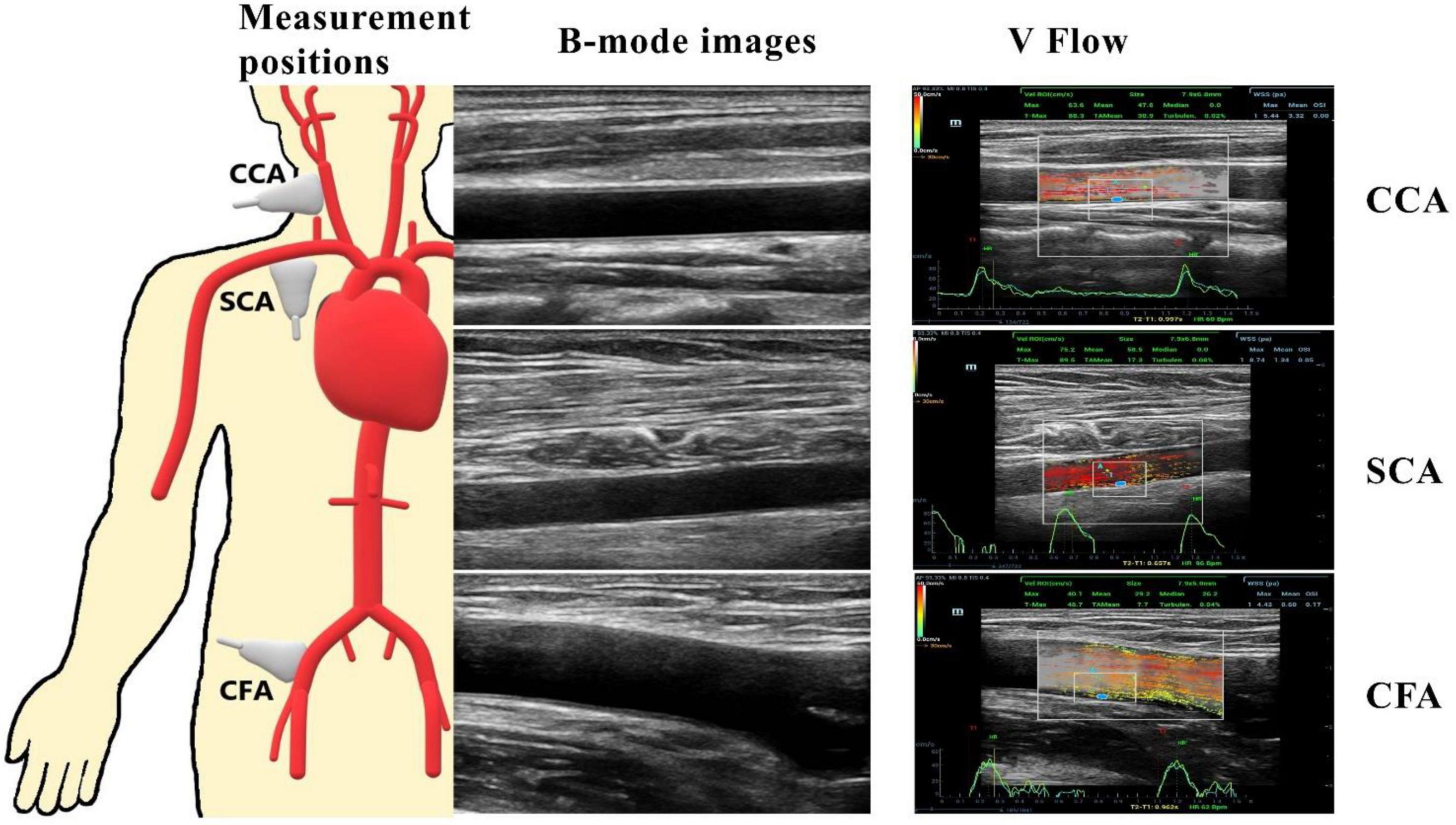
Frontiers Investigation on the differences of hemodynamics in normal common carotid, subclavian, and common femoral arteries using the vector flow technique
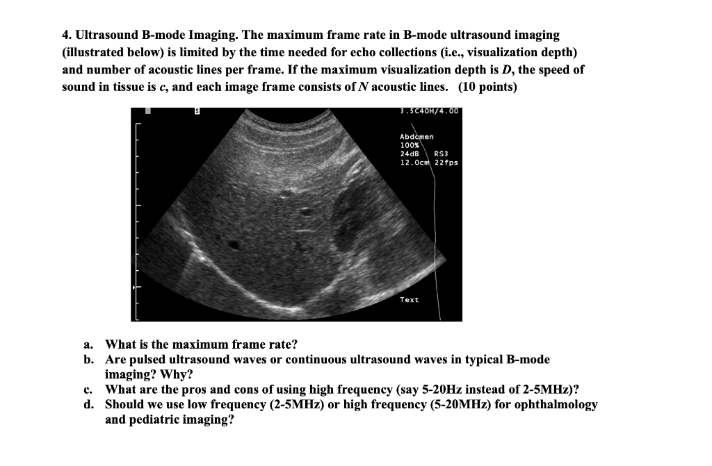
Solved Are pulsed ultrasound waves or continuous ultrasound
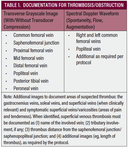
Duplex Ultrasound Technical Considerations for Lower Extremity Venous Disease - Endovascular Today
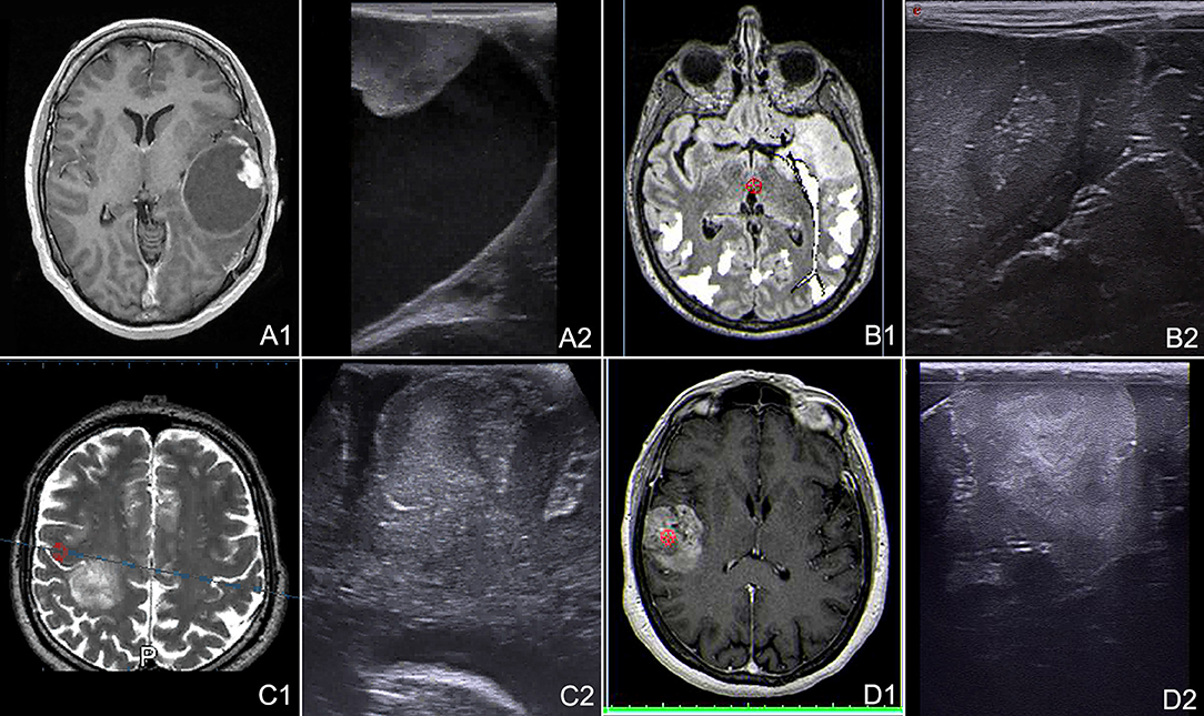
Frontiers Advanced Ultrasound Imaging in Glioma Surgery: Beyond Gray-Scale B-mode

Figure 1 from Deep Learning in RF Sub-sampled B-mode Ultrasound

Jong Chul YE, Ph. D.

Physics of Ultrasound! – Critical Care Northampton

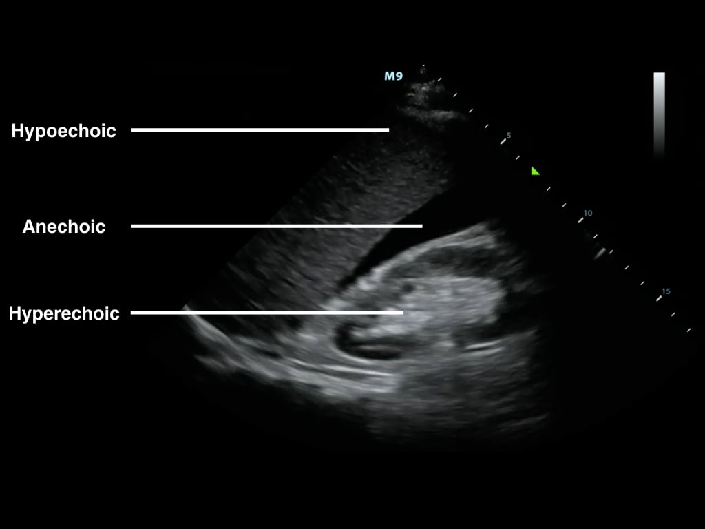
Grey scale imaging (ultrasound), Radiology Reference Article

PDF) Deep Learning in RF Sub-sampled B-mode Ultrasound Imaging

Ultrasound Doppler-guided real-time navigation of a magnetic microswarm for active endovascular delivery
Ultrasound Physics and Technical Facts for the Beginner
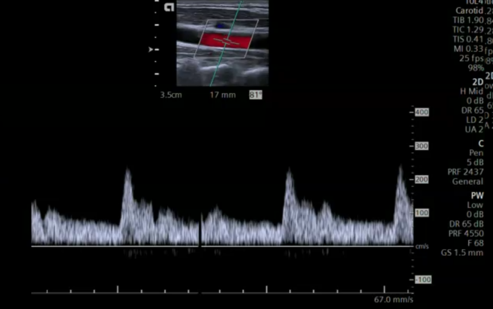
5. Color Doppler imaging of the carotid arteries