Most clots retrieved from patients with acute ischemic stroke are ‘red’ in color. ‘White’ clots represent a less common entity and their histological …
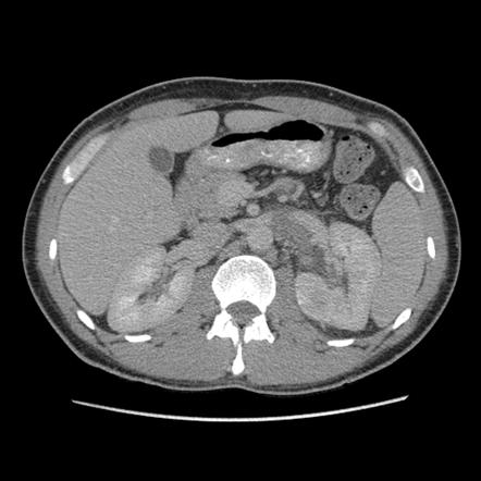
Renal vein thrombosis, Radiology Reference Article

Comprehensive and Structured 3-month Stroke Follow-up Using the

Thrombus composition and thrombolysis resistance in stroke - Research and Practice in Thrombosis and Haemostasis

Characterization of the 'White' Appearing Clots that Cause Acute Ischemic Stroke - ScienceDirect
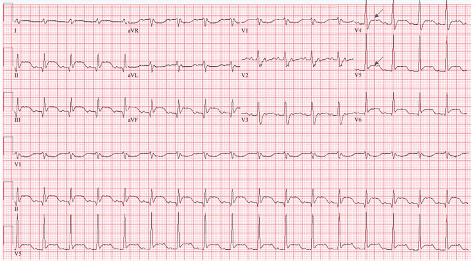
An unexpected turn: A 71-year-old man with myocardial infarction

Antithrombotic Therapy for Primary and Secondary Prevention of

Bilateral Large Vessel Occlusion Causing Massive Ischemic Stroke

Characterization of the 'White' Appearing Clots that Cause Acute Ischemic Stroke - ScienceDirect

Gross specimen (a1, b1), H&E staining (a2, b2) and clot analysis (a3
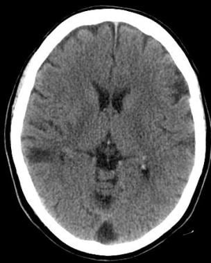
Stroke Imaging: Practice Essentials, Computed Tomography, Magnetic Resonance Imaging

REVIEW: “ISCHEMIC STROKE: From Fibrinolysis to Functional Recovery

Predictive value of clot imaging in acute ischemic stroke: A systematic review of artificial intelligence and conventional studies - ScienceDirect

Central Pulmonary Embolism Detected on a Chest X-Ray: A Case Report - Journal of the Belgian Society of Radiology

JCM, Free Full-Text

:max_bytes(150000):strip_icc()/VWH-ZoeHansen-PotentialSignsofMiscarriage-Standard-b964332362dc45fa806d18a4ad6a221d.jpg)